It was a quiet Saturday morning when the owners of Little Jack, a 9-year-old Maltese, knew something was wrong. When he woke up, Little Jack let out a yelp, and seemed to be in distress. His owners rushed him to their local veterinarian who discovered that Little Jack was as yellow as a banana! Jokes aside, this can be a serious life-threatening sign that indicates a major dysfunction of the liver and is known as jaundice or icterus. Jack was immediately hospitalized for investigations and supportive treatment.
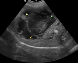
After stabilization and management of Jack’s pain, the primary care veterinarian decided to refer him to our specialists in Central London for advanced testing and treatment. Upon arrival to London Vet Specialists, Little Jack first met Dr David Neilson, our anaesthetist and pain management expert. David rapidly went about trying to make Jack as comfortable as possible, while Dr Ian Jones our specialist in diagnostic imaging, performed an abdominal ultrasound of Jack’s belly. Jack was diagnosed with a biliary mucocele which means that his bile was getting too thick inside of the gall bladder and his body was not able to release it into the small intestines. Normal bile flow is critical for digestion. Below we can see an image of the ultrasound performed. The arrows outline the thick bile inside the gall bladder. Normally the inside of the gall bladder should appear uniformly black (indicating liquid) on an ultrasound, not filled with white and grey (indicating thick debris).
This is a well-known condition in animals and humans and once the gall bladder is blocked the treatment often means surgery to remove the gall bladder. Specialist surgeon, Dr Janet McClaran and the rest of the surgical team were ready for the emergency procedure, and in within hours, Little Jack had his gall bladder removed and was comfortably recuperating.
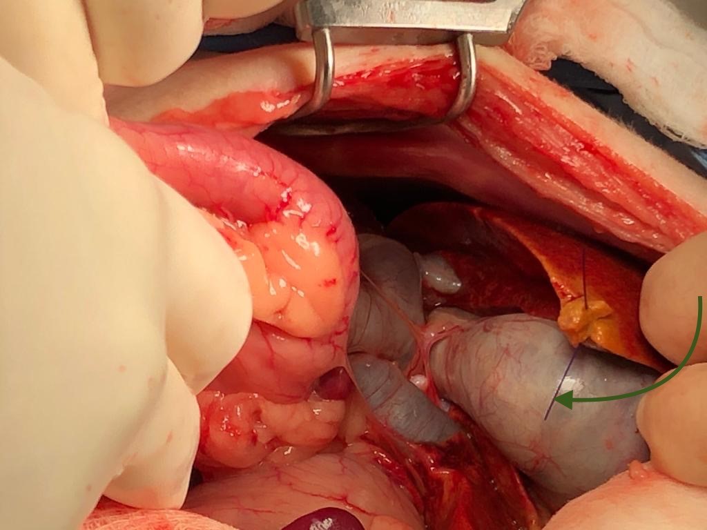
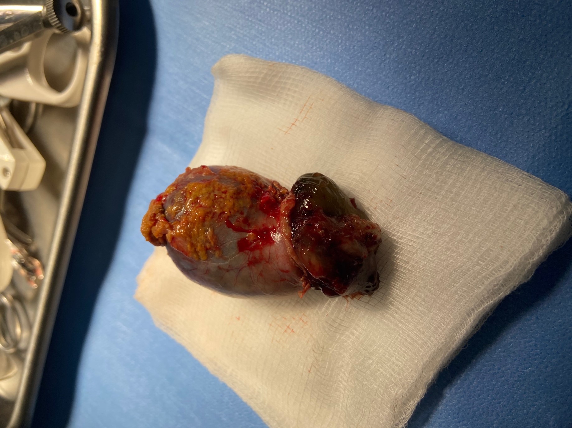
If you are not squeamish, below are some pictures of the surgery. The first image has a green arrow pointed to the very enlarged white gall bladder near the liver (the liver has a suture in place from a biopsy). The second picture below shows the gall bladder, full of debris, after being removed from Jack.
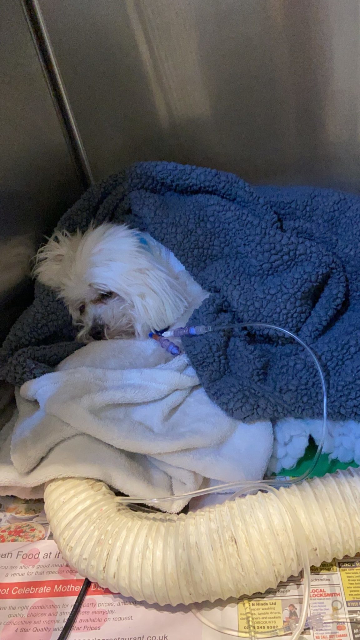
Several tubes and lines were attached to Little Jack, allowing us to measure blood pressure, take blood samples, and provide him with medications and pain killers. Additionally, a feeding tube was placed to administer food straight into his stomach. Here is Jack recovering from surgery.
This surgery, called a cholecystectomy, has a high rate of complications in dogs. Dogs with gall bladder issues can be slow to heal and have trouble processing the anaesthetics, but Little Jack proved to all of us that he is a Little Warrior, and he responded brilliantly from surgery. He remained in hospital for several days following surgery for pain control and monitoring and then he continued recuperating at home with the love of his family. He has made a full recovery with no signs of yellow or jaundice. His care has been transferred to our liver expert, medic Dr. Ellie Mardell, who will manage his follow up care (including diet and medications to strengthen his liver) in partnership with his primary care veterinarian.