Muffin is a 3-year-old lovely little pug, that initially presented to her primary care veterinarian following a few days of tummy upset, having some vomiting, diarrhoea and turning down her supper.
She was admitted to hospital, and while she responded to the initial supportive care, including intravenous fluids and anti-nausea medications, she suddenly took a turn for the worse. She started to have difficulty breathing, and anaemia (a low red blood cell count). Pneumonia was suspected.
A chest radiograph (xray) showed fluid in her chest, and transfer to the London Vet Specialists (LVS) team of experts was elected. She was swiftly cared for, oxygen provided, and a blood transfusion elected to make her stronger.
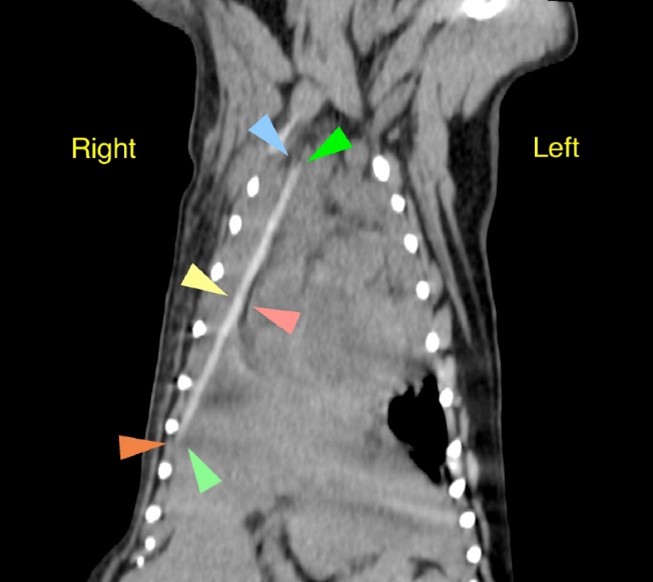
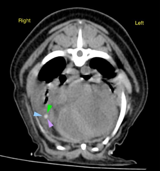
A CT scan (a three-dimensional xray that provides superior detail of the chest cavity) was performed by our imaging specialist, Dr. Ian Jones. The result was definitely unexpected! A linear object consistent with a piece of wood or stick was found in the right side of her thorax! See the CT images below with arrow heads highlighting the offending object.
Muffin was swiftly transferred to the care of the LVS surgical team (including Dr Janet Mcclaran, surgical intern Dr Fabio Esposito, surgical nurse Holly Brown, and the anaesthesia team led by specialist Dr David Neilson) for emergency intervention and the delicate operation to remove the stick.
Foreign material such as sticks or kabobs may enter the chest cavity from an external penetrating injury (such as running into a tree!) but more commonly, foreign material migrates into the chest, through the diaphragm, after a stick is ingested and then perforates (makes a hole) tunnelling out of the gastrointestinal tract. . In Muffin’s case surgery was initiated in her abdomen and a small penetrating wound was found in the duodenum (small intestine) where the stick exited and then crossed her diaphragm and entered the chest cavity.
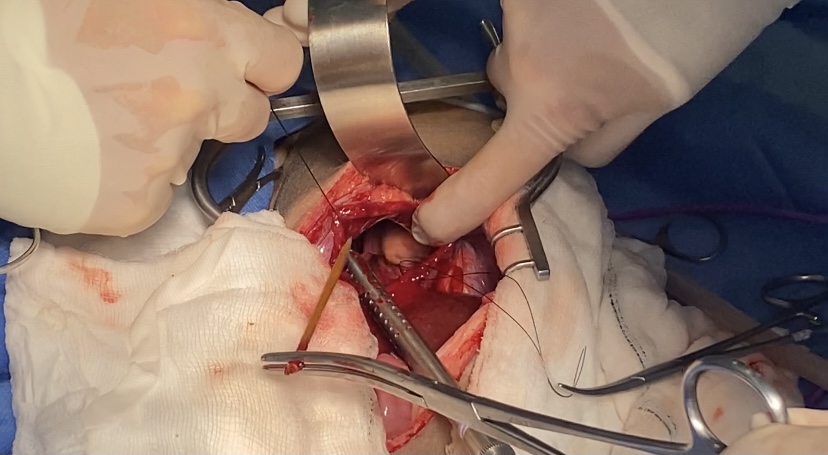
A window into the chest was created through the diaphragmatic and the chest cavity was carefully inspected. As suspected, a wooden skewer was found between her right lung lobes, but very luckily for Muffin, it had not damaged her lungs, heart, or any of the major blood vessels such as the aorta or vena cava. See photos of the kabob stick being removed from Muffin, below.
Muffin showed all her strength during the recovery and after several days of close monitoring and recovery with the LVS team, she was able to return to her family after just few days of hospitalization.
The LVS staff was delighted to see Muffin again for her last post-operative check (see photo below). She looked very energetic, and back to her happy pug self! We wish her all the best and have advised against any future late night cheeky kabobs!