A 7-year-old Domestic Shorthair female neutered was referred to London Vet Specialists (LVS) due to pyrexia (39.2 degrees C), anorexia and lethargy. Blood tests revealed hematocrit of 23% and a high level of creatinine (172 μmol/L). On clinical examination, she was uncomfortable on palpation, especially over the kidneys.
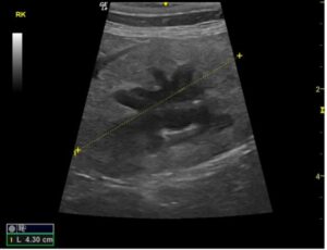
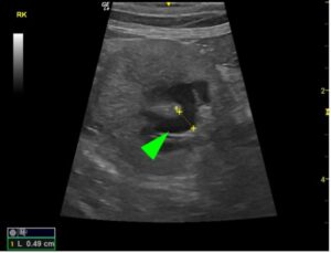
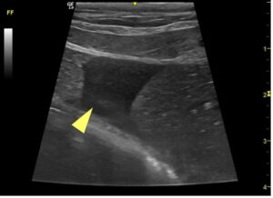
An abdominal ultrasound was performed (see images), and a marked hyperechoic cortical part of the right kidney (RK) and a moderate distention of the pelvis (0.5 cm) (green arrowhead) with normal size of the kidney were identified.
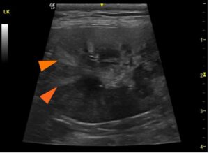
The left kidney (LK) was also normal size, but multiple hyperechoic wedge-shape areas on the cortical part (orange arrowheads) and slight loss of corticomedullary differentiation were also observed.
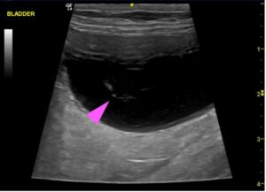
The bladder was moderate distended with the presence of small amount of hyperechoic mobile content (pink arrowhead).
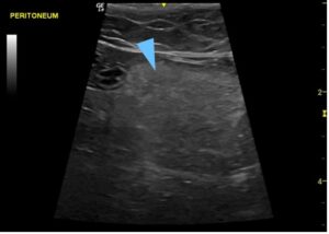
A small amount of free anechoic peritoneal fluid (yellow arrowhead), peri-renal fluid around the right kidney and hyperechoic peri-renal fat (blue arrowhead) around the right kidney were also visible.
The main differential diagnosis was pyelonephritis of the right kidney, multiple left kidney infarcts, with signs of chronic kidney disease and urinary sediment. A sterile urine sample was taken by US-guided cystocentesis for culture and sensitivity analysis, and Escherichia Coli sensitive to amoxycillin/clavulanic acid was isolated. Treatment was implemented and the patient recovered from her issue.
US-guided cystocentesis is one of the most common techniques used in small animals to take sterile urine samples. This technique has lower risk than blind cystocentesis, because the bladder can be visualized directly on the ultrasound and while the needle is being inserted into the bladder to take the sample, avoiding damage of other organs. Cystocentesis is also very useful when a urine infection is suspected, because the urine sample is directly collected from the bladder, avoiding the possible contamination when the sample is taken with spontaneous miction. Referral was crucial in this case, to safely acquire images, samples, and get an early diagnosis for the patient. If you have any questions about referral to our internal medicine or diagnostic imaging team, please contact us.