Rosie is a lovely 15-year-old collie cross that presented to London Vet Specialists with a huge, grapefruit sized swelling under her tail. Her owner noticed that something was not quite right. Rosie was having problems urinating, straining to defaecate and overall seemed bothered by her rear end. Her primary care veterinarians discovered a lump of her perineal region (the area between her anus and vagina). They decided to call on the team of specialists at LVS to help sort out the problem. This region can be very difficult to examine, and what is visible from the outside is often just “the tip of the iceberg.” A CT scan (a 3D X- ray) can provide much more detailed information compared to traditional X-rays. This type of scanner is only available at select locations, and LVS is the only facility in London to have a scanner that is run by a specialist in Diagnostic Imaging, Dr Ian Jones. CT scans help to plan surgery by providing a visual map detailing the extent of tumours and helps us to determine if tumours have spread (metastasized) to any distant locations in the body- just like in people!
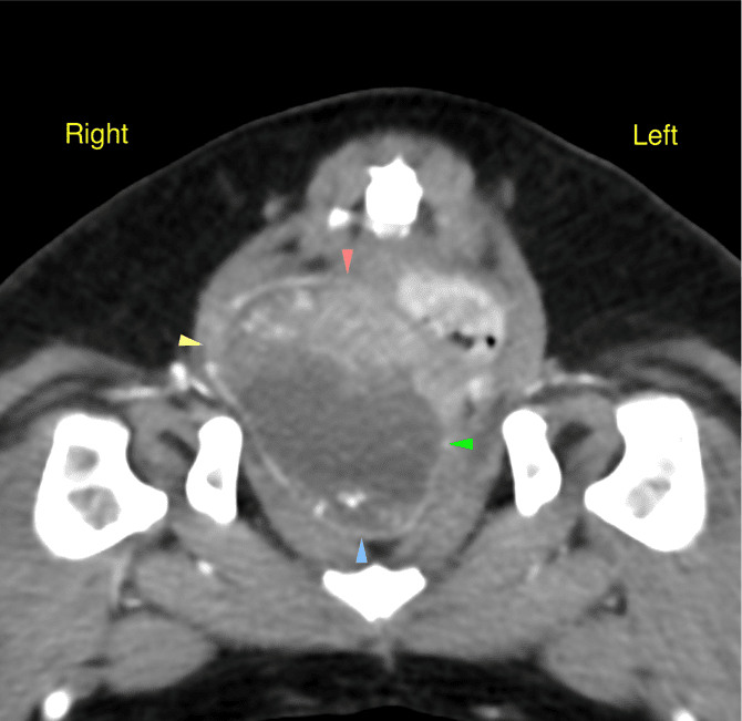
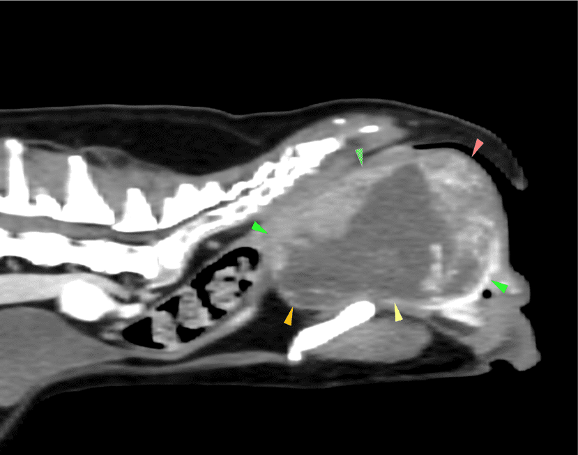
Rosie’s CT scan revealed a huge mass growing from her vagina, as you can see in the images below (arrows outlining the mass). This tumour was compressing her urethra and colon causing her much discomfort, especially when urinating and defaecating.
This was a delicate operation to perform as there is limited access to the region and there was a possibility she could require a blood transfusion (as vaginal tissue can bleed profusely.) Surgery in elderly patients comes with more risk than in younger patients. All the pre-operative testing proved Rosie to be a good candidate for surgery and “age is not a disease!” Luckily for Rosie, we have a specialist in Anaesthesia, Dr David Neilson, to monitor her procedure and recovery. Specialists undergo years of advanced training and examining following graduating from veterinary school, to provide the most state-of-the-art care for animals.
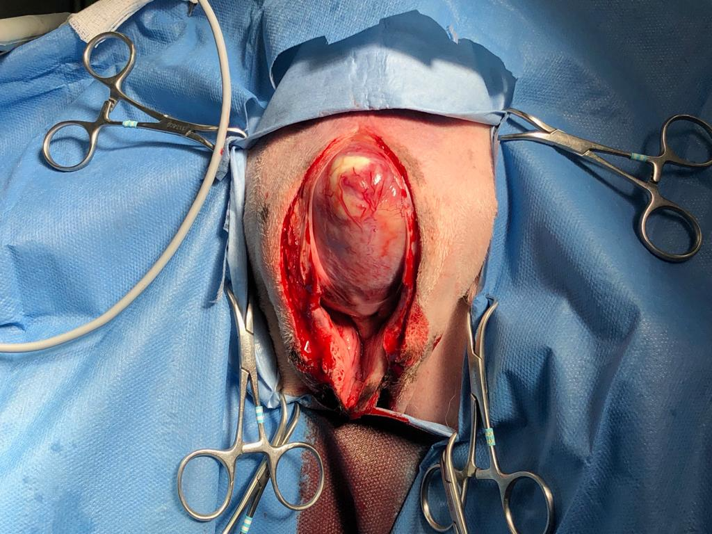
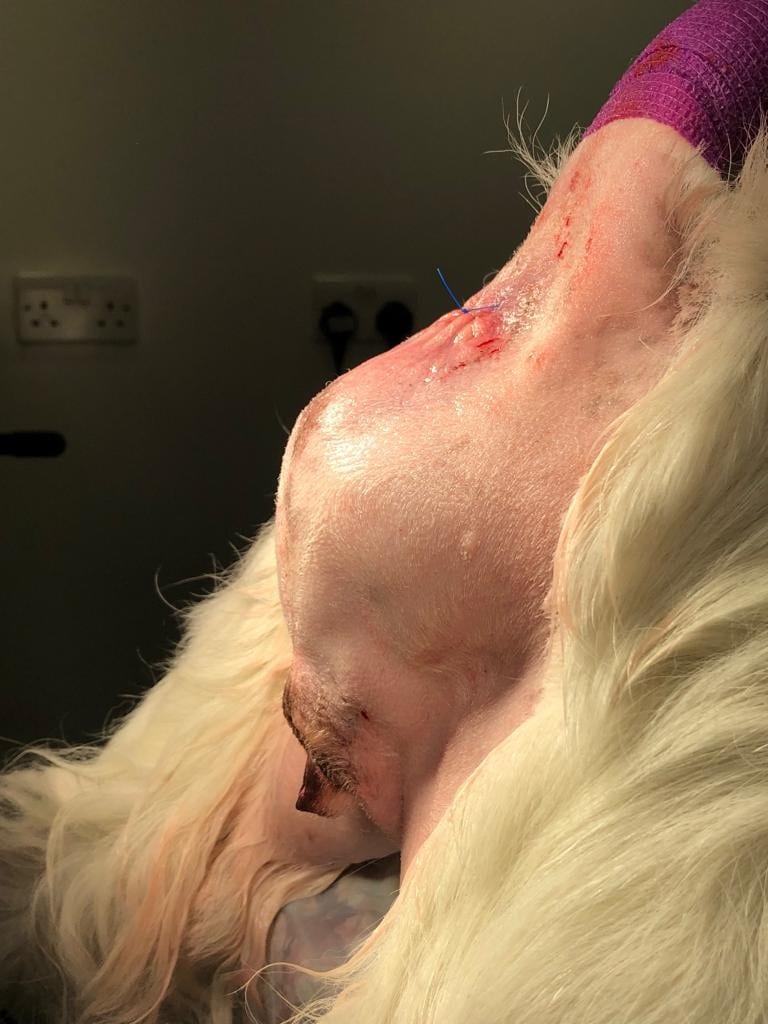
Surgery was performed by our soft tissue specialist, Dr. Janet Kovak McClaran, who has years of expertise in removing tumours. The large vaginal mass was accessed via an incision underneath her tail. The image from surgery is below (please be aware it is a bit graphic and it is not for sensitive people, so if you are squeamish, stop here!)

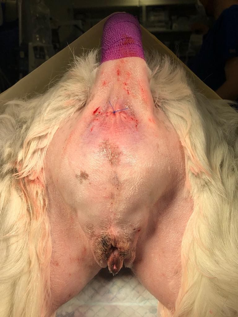
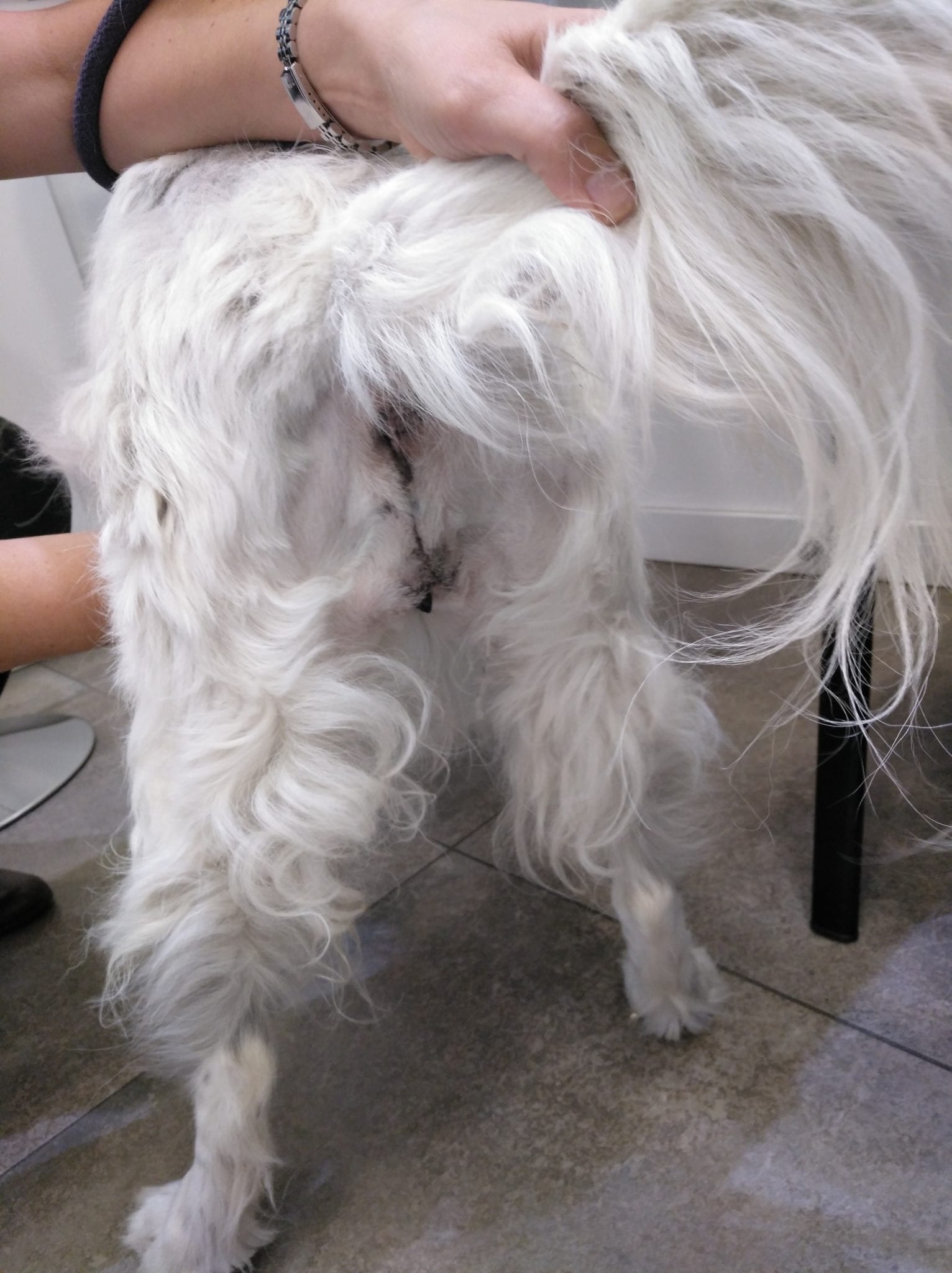
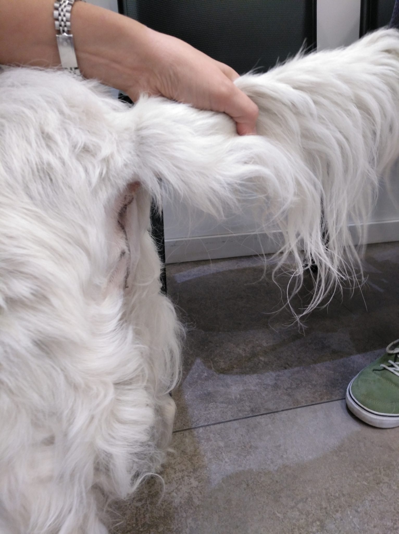
Below are some “before and after” pictures. On the left the large mass is visible under her tail and on the right are the photos taken 2 weeks following her surgery. Final biopsy revealed the great news that this was a very slow growing tumour called a leiomyoma. It was completely removed and unlikely to cause Rosie any problems in the future.
Now, thanks to the LVS team of superheroes, Rosie is happy, full of energy and has a newfound spring in her step.
